
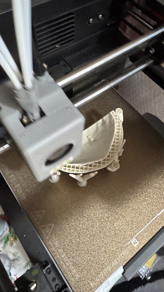
3D printing in medicine lets surgeons hold in their hands an exact model of the organ they are about to operate on – before they even enter the operating room. This is no longer science fiction but a reality in several hospitals in North Macedonia, doctors reveal in the latest issue of Medici, the journal of the Medical Chamber of North Macedonia.
How 3D Printing in Medicine Works
Using computed tomography (CT) or magnetic resonance imaging (MRI) scans, a team of doctors and engineers creates a digital and then a physical model of the specific patient’s organ, bone or blood vessel. Unlike an ordinary scan – a two-dimensional image on a screen – the model gives a true spatial representation, at actual size.
The process usually looks like this:
- Imaging – the patient has a CT or MRI scan, just as for any other operation.
- Segmentation – the organs, bones and blood vessels relevant to the operation are isolated from the scans.
- Digital model – a precise 3D model is created, which the surgeon reviews and approves.
- 3D printing – the model is printed at actual size, and its accuracy is then verified.
- Planning – the surgeon uses the model to plan and “rehearse” the operation and to explain it to the patient.
Real Cases from North Macedonia
In Medici, doctors describe several striking cases: a complete nasal reconstruction in a patient after a malignant disease, a skull reconstruction in a young man after a traffic accident, and even a heart model of a two-month-old baby with a severe heart anomaly. In all of these cases, the 3D model was not just an aid – it also influenced the decision on how the operation would be performed.
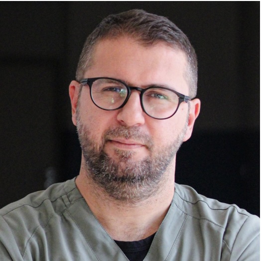
“Creating 3D models enables proper planning, which reduces the risk of surgical errors while increasing effectiveness.” – Asst. Dr. Valon Saliu, digestive surgeon, for Medici
The 3DLoom team also publishes its work in the professional literature – for example, a case report in the Journal of Surgical Case Reports (Oxford Academic) on how a 3D printed model helped plan a complex operation on the pancreas and spleen. You can find more cases on our Press & Publications page.
Where 3D Printing in Medicine Brings the Greatest Benefit
- Abdominal surgery – liver and pancreatic operations where the tumor lies close to major blood vessels.
- Reconstructive surgery – reconstruction of the skull, nose and face, where the goal is both function and symmetry.
- Cardiac surgery – complex heart anomalies, especially in children.
- Orthopedics and traumatology – complex fractures and bone deformities.
- Medical education – 3D printed bone replicas for students and residents.
What This Means for Patients
Although this technology has been standard in developed countries for two decades, here it is still in its early stages. That is exactly why 3DLoom is working to make the technology accessible to more hospitals and more patients across the country – without every institution having to build its own expensive laboratory.
For patients, this means operations are becoming more predictable, more precise and safer. And behind every successful outcome lies not only the surgeon’s skill, but also hours of preparation by the teams developing the 3D technology.
Frequently Asked Questions
Is 3D printing in medicine safe?
Yes. The model is used outside the patient’s body as a planning aid, and every model is checked by the surgical team before use.
Is this an experimental treatment?
No. 3D models are a planning aid for already established surgical procedures – the operation itself is performed in the standard way.
Does every operation require a 3D model?
No. The greatest benefit is in complex, rare or anatomically unusual cases, where the surgeon assesses whether the model will help with planning.
Read more about how 3DLoom works and how 3D printing in medicine can help in your case too – or contact us.
Source: Medici, journal of the Medical Chamber of the Republic of North Macedonia, VOX 131, June 2026.
 English
 Macedonian